 |
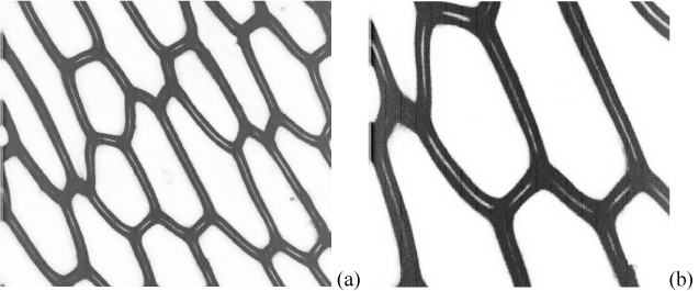
| Figure 4: Typical acoustic images of onion epidermal walls at a frequency of 600 MHz. The white lines are identified as cuticle structures located between adjacent cell walls making up the risers in the “shoe-box” structure. In (a) the scan width is 1 mm, whereas in (b) the scan width is 0.5 mm. |