 |
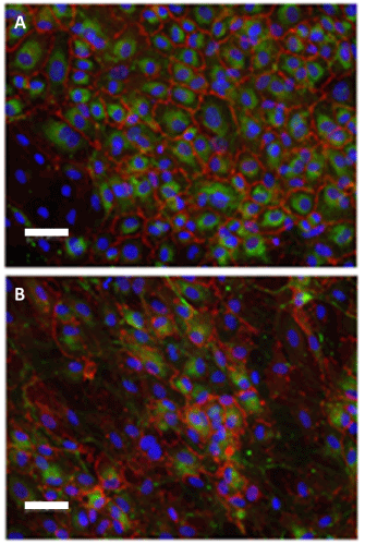
| Figure 2: Endothelial cells cultured on PTFE membranes and incubated with whole blood. A–cells after 36 hours of static culture. B–cells after 36 hours of dynamic culture. Immunofluorescence staining against von Willebrand factor (green) and CD31 (red). Scale bar–50 μm. |