|
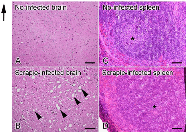
| Figure 1: Light micrograph images of mouse brains (A, B) and spleens (C, D) 155 days post-inoculation stained with hematoxylin and eosin. (A, B) Many vacuoles are observed throughout all areas of the brain of a scrapie-infected mouse (arrowheads), while no vacuole is seen in the brain of a no-infected mouse. (C, D) Lymph nodules containing germinal center (asterisk) are observed in the spleens of both a no-infected mouse and a scrapie-infected mouse. The germinal center of a scrapie-infected mouse is diffusely large, suggesting an immune response to the infection. Bars=100 μm. |