|
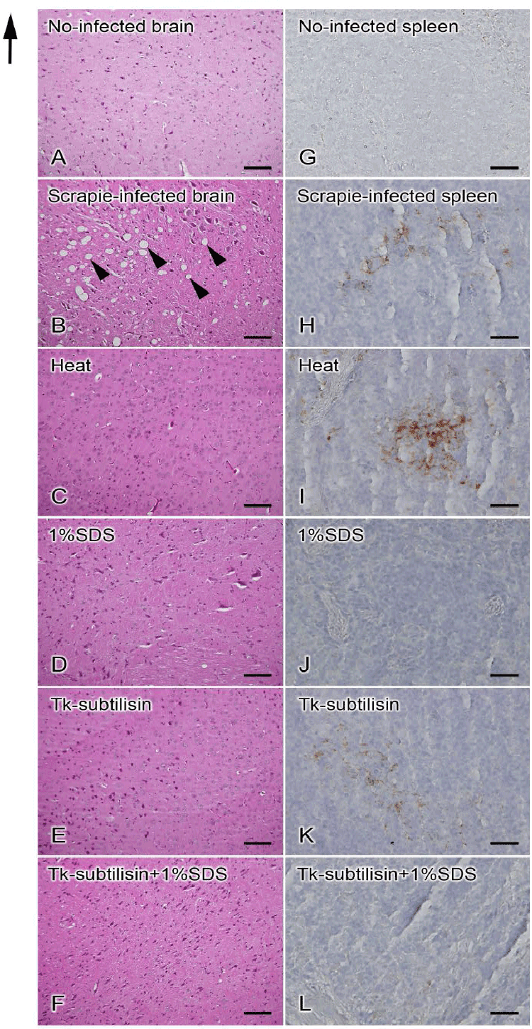
| Figure 2: Light micrograph images of mouse brains (A-F) and spleens (G-L) 155 days post-inoculation. (A-F) Neuropil vacuolation is observed throughout all areas of the brain of (B) a scrapie-infected mouse (arrowheads), whereas no vacuolation is seen in the brain of (A) a no-infected mouse, (C) a mouse inoculated with SBH incubated at 100°C, (D) a mouse inoculated with SBH incubated at 100°C with 1% SDS, (E) a mouse inoculated with SBH digested with Tk-subtilisin, and (F) a mouse inoculated with SBH incubated with Tk-subtilisin and 1% SDS. (G-L) Light micrograph images showing the localization of PrP in the spleen of (G) a no-infected mouse, (H) a scrapieinfected mouse, (I) a mouse inoculated with SBH incubated at 100°C, (J) a mouse inoculated with SBH incubated at 100°C with 1% SDS, (K) a mouse inoculated with SBH digested with Tk-subtilisin, and (L) a mouse inoculated with SBH incubated with Tk-subtilisin and 1% SDS. Immunoreactivity for PrP (brown signal) is detected around the center of the lymph nodule in the spleen of (H) a scrapie-infected mouse, (I) a mouse inoculated with SBH incubated at 100°C and (K) a mouse inoculated with SBH digested with Tksubtilisin. No labeling for PrP is observed in the lymph nodules (G, J, L). Bars: A-F=100 μm; G-L=50 μm. |