|
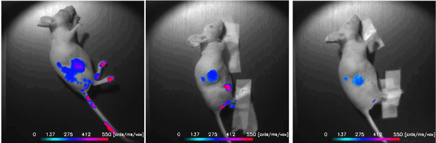
| Figure 4: In vivo NIRF Imaging of BI-10AF750. Three mice were i.v. injected with 80 װg of the BI-10AF750 MI probe, and NIR fluorescence images were acquired 6 hours post injection. A threshold was applied to better depict the tumors. Signals in the kidneys and bladder indicate renal filtration. No signal was observed in the liver at this time. An indication of tumor cell growth along the subcutaneous needle injection was observed in two mice (left and right). |