|
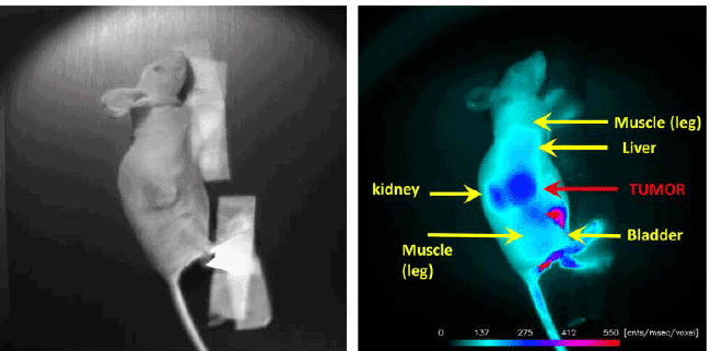
| Figure 5: NIR Optical Images of BI-10AF750 Molecular Imaging (MI) Probe. (Left) Displayed is the bright field image of a mouse with an ovarian tumor on its flank, and (Right) the corresponding fluorescence image 6 hours post-injection of the BI-10AF750 MI probe. The arrows depict the anatomical features of the mouse. |