|
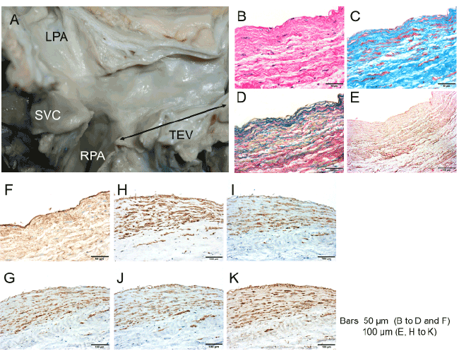
| Figure 1: Macroscopic and microscopic findings of human tissue-engineered vasculature (TEV) Panel A shows the macroscopic appearance of the TEV (LPA, left pulmonary artery; RPA, right pulmonary artery; TEV, black double arrowheads). Panels B, C, D, and E show hematoxylin and eosin staining, Masson’s trichrome staining, Victoria blue–van Gieson staining, and modified von Kossa staining, respectively. Panel F shows factor VIII-positive cells (brown), which represent the endothelial cells of the TEV. Panels G, H, I, J, and K show cells positive for alpha smooth muscle actin, myosin heavy chain, smooth muscle myosin heavy chain 1, smooth muscle myosin heavy chain 2, and calponin (brown), respectively. |