 |
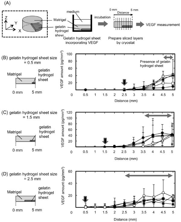
| Figure 3: Time profiles of VEGF release from the gelatin hydrogel sheets incorporating 3 ng of VEGF. (A) Schematic illustration of diffusion patterns of VEGF from gelatin hydrogel sheet incorporating VEGF in the X-axis direction of Matrigel. (B-D) Amount of VEGF in the X-axis direction from the left end of GML. The size of gelatin hydrogel sheet was 0.5 (B), 1.5 (C), and 2.5 mm (D). The incubation time was 0 (○), 15 (Δ), and 30 min (□) or 1 (●), 3 hr (▲). |