|
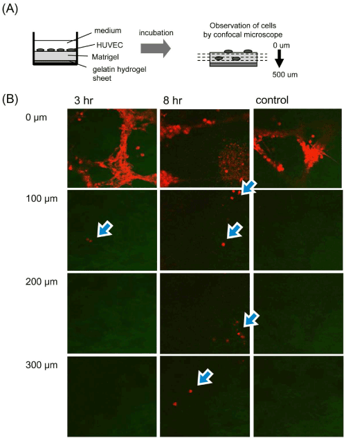
| Figure 5: Migration patterns of HUVEC into Matrigel by VEGF released from gelatin hydrogel sheets. (A) Schematic illustration of cells observation in the Z-axis direction of Matrigel through VEGF release from the gelatin hydrogel sheet incorporating VEGF. (B) Fluoresent microscopic pictures of Matrigel layers after 3 and 8 hr incubation. Cell migration was evaluated by VEGF-free gelatin hydrogel sheets after 8 hr incubation (control). Arrows indicate cells migrated into Matrigel. |