|
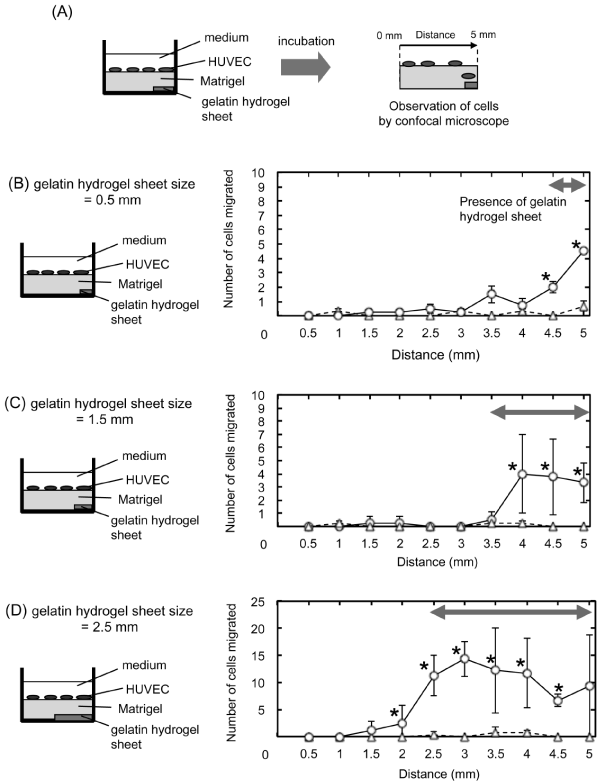
| Figure 7: Migration patterns of HUVEC into Matrigel by VEGF released from gelatin hydrogel sheets incorporating 670 pg of VEGF. (A) Schematic illustration of the cells observation in the X-direction of Matrigel through VEGF release from the gelatin hydrogel sheet incorporating VEGF. (B-D) Amount of cells migrated in the X-axis direction of Matrigel after 8 hr incubation. The gelatin hydrogel sheet size was 0.5 (B), 1.5 (C) or 2.5 mm (D). Each gelatin hydrogel sheet incorporates VEGF (○) or PBS (Δ). *P<0.05; significance against the number of cells migrating of using PBS incorporating gelatin hydrogel sheet. |