|
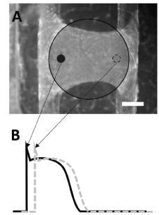
| Figure 4: A Potential Artifact of Action Potential Profile. (A) Engineered heart tissues loaded with FluoVolt and schematically illustrated two different spots within an area of optical detection illuminated by a diffused excitation focus. Bar = 1mm (B) Delayed action potential profiles from two spots in (A). |