 |
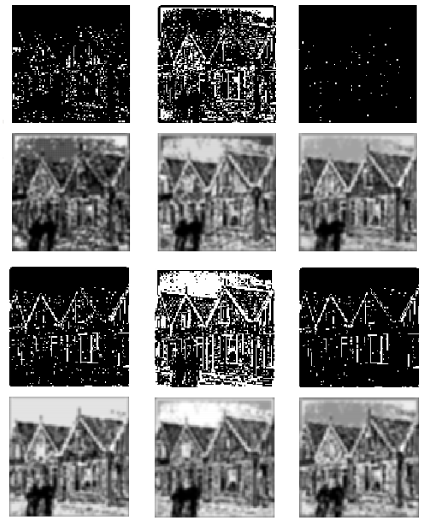
| Figure 6: Simulation of phosphene-based reconstruction of an image using the STEMLIS method. First and third rows of images: stimulation sequence (ganglion outputs obtained from artificial retina model). Second and fourth rows of images: phosphene images reconstructed using the STEMLIS method. Frames 5, 8, 11, 14, 18, and 22 were selected from 30 ms of total sequence for this illustration. |