 |
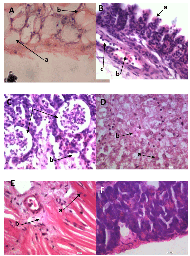
| Figure 5: Histopathological section of A: Skin; (a) necrosis in the dermal layer (b) hypertrophy in immersion group with (H&E stain100X). B: Gill; (a) hyperplasia in the secondary lamellae (b) leukocytic infiltration(c)dilation of the central venous sinus (H&E stain100X) C: Kidney; (a) degenerative changes in glomerular epithelium (b) inflammatory cells (H&E stain 100X) D: liver; (a) hepatocytes vacuolar degeneration (b) inflammatory cells (H&E stain 100X) E: Muscles; (a) mild edema (b) focal hyaline degenerationF: Spleen; hyperplasia in the lymph follicles. |