|
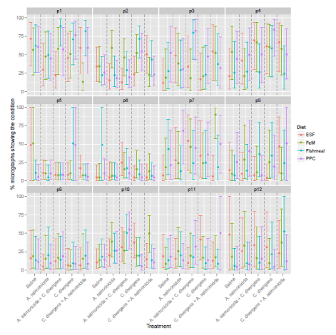
| Figure 2: Expected % of 10 micrographs per fish showing the specific condition with 95% confidence intervals as indicated by the fitted statistical model. Subplots denote conditions p1-p12 (denoted in strip text). Colors represent background diets. The parameter for which the micrographs were scored for are: mitochondria (healthy [P1] or unhealthy [P12]), mitochondrial anchorage (P2), edema (P3), vacuolization (P4), presence of rodlet cells (P5), bacteria-like particles (P6), inter-epithelial lipid storage (P7), terminal web (P8), intraepithelial leukocytes (IEL) (P9), damaged microvilli (P10) and the presence of cell debris in the lumen (P11). |