|
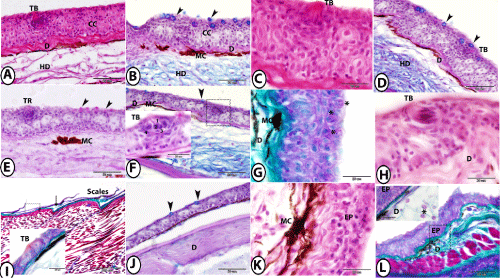
| Figure 2: Organization of skin of red tail shark at different body regions. A and B: The skin of the lower lip stained by HE and PAS-AB-HX respectively showing taste bud (TB), club cells (CC), mucous cells (arrowheads) and melanocytes (MC) at the dermis (D) followed by hypodermis (HD). C and D: The skin of the upper lip stained by HE and PAS-AB-HX respectively showing taste bud (TB), mucous cells (arrowheads) and thin dermis (D) followed by hypodermis (HD). E: The skin of the snout stained by PAS-AB-HX showing PAS positive mucous cells (arrowheads), tuberous organ (TR) and melanocytes (MC). F: The skin of the operculum stained by PAS-AB-HX showing mucus cells (arrowhead), taste bud (square, TB, inserted figure) that consisted of sensory cells (1), supporting cells (2), basal cells (3) and mentle cells (4). Note, melanocytes (MC) on the dermis(D). G and H: The skin of the dorsum of the head stained by Crossmon's Trichrome and HE respectively showing rodlet cells (asterisk), taste bud (TB), melanocytes (MC) on the dermis (D) of collagenous fibers. I: The skin of the trunk stained by Crossmon's Trichrome showing scale pockets (arrow), taste bud (square, TB, inserted figure). J: Tigher magnification of square of figure. I showing the skin of trunk stained by PAS-AB-HX indicates positive stained mucous cells (arrowheads). Note parallel bundles of collagenous fibers in the dermis (D). K and L: The skin of the tail stained by HE and Crossmon's Trichrome respectively showing melanocytes (MC) in the deeper layers of epidermis (EP). The dermis (D, inserted figure in L) contained many rodlet cells (asterisk). |