 |
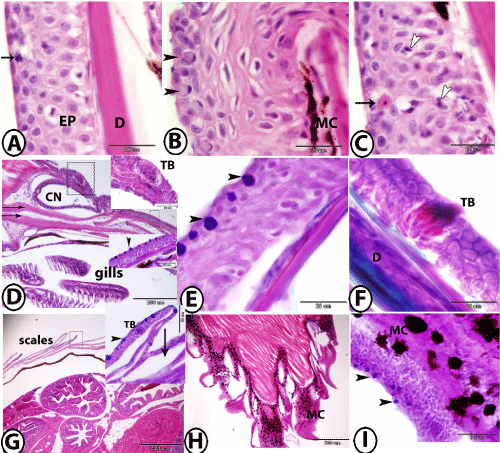
| Figure 3: Organization of skin of guppy at different body region. A: The skin of the lower lip stained by HE showing mitotic cells (arrow) in stratified squamous epidermis (EP), followed by the dermis (D). B: The skin of the upper lip stained by HE showing mucous cells (arrowheads) and melanocytes (MC). C: The skin of the snout stained by HE showing eosinophilic granular cells (arrow) and lymphocytes (arrowheads). D: The skin of the operculum stained by HE showing taste bud (large square, TB). The small square indicates mucous cells (arrowhead) in the epidermis of the inserted figure stained by PAS-AB-HX. Note presence of canal neuromast (CN) surrounded by osteoid tissues (arrows). E and F: The skin of the dorsum of the head stained by PAS-AB-HX and Crossmon's Trichrome respectively showing mucous cells (arrowheads) and taste bud (TB). Note the dermis (D) is formed of collagenous fibers. G: The skin of the trunk stained by HE showing scale pockets (arrow). The square indicated the inserted figure stained with PAS-AB-HX showing taste bud (TB) and mucous cells (arrowhead). H and I: The skin of the tail stained by HE and PAS-AB-HX respectively showing numerous black melanocytes (MC) and many mucous cells (arrowheads). |