|
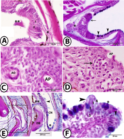
| Figure 5: The lateral line system of guppy. A: the ampullary organ is formed of sensory cells (1), supporting cells (2) and covered by cupula (2 asteriks). Note presence of nerve fibers (asterisk) basally. B: ampullary organ stained with PAS-AB-HX showing AB-positive apical border of supporting cells (arrow), basal bundle of nerve fibers (asterik). Note that the ampullary channel contained PAS-AB positive mucous cells (arrowheads). C: a horizontal skin section of the epidermis stained by HE showing an ampullary pore (AP) in close proximity to an ampullary organ (AO). D: a vertical skin section stained with HE showing cross section of the ampullary organ (arrow) under an ampullary pore (AP). E: canal neuromast (square, inserted figure) stained with PAS-AB-HX showing the dermal bone (DB) that surrounded the canal neuromast (CN), which lined by sensory cells (arrowhead), AB-positive mucous cells (asterisk). Note presence of nerve fibers (two asterisks) below the neuromast. F: Superficial neuromasts (arrowheads) stained by PAS-AB- HX is formed of sensory cells (1), supporting cells (2), basal cells (3) and surrounded by mentle cells (4). Note presence of mucous cells (asterisk) near neuromasts. |