|
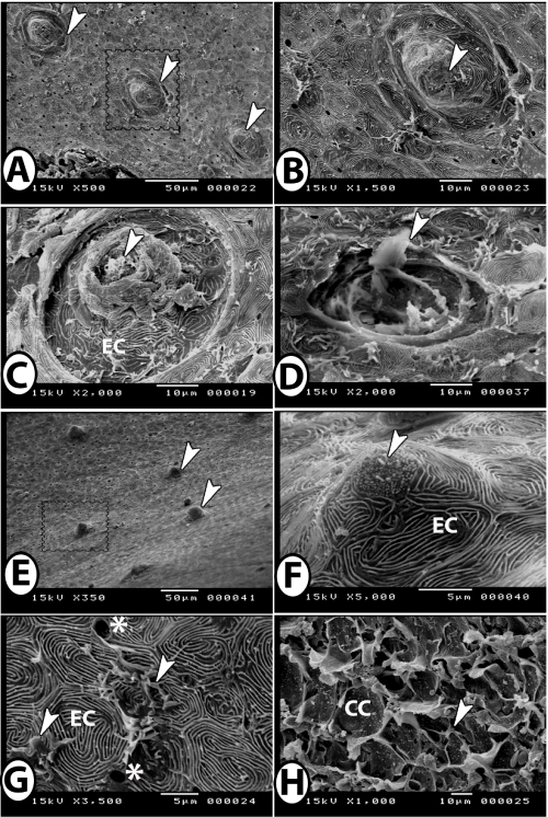
| Figure 8: Neuromasts and taste buds of red tail shark. A: Superficial neuromasts arranged in rows (arrowheads). B: Higher magnification of the square in Figure A showing superficial neuromast (arrowhead). C: Ring like pattern of neuromast formed of epidermal cells (EC) with hairs from sensory cells at their apex (arrowhead). D: Superficial neuromast with attached cupula (arrowhead). E: Epidermal elevations (arrowheads, square). F: Conical non-keratinized protuberance of epidermal cells (EC) with a taste bud at a summit (arrowhead). G: surface of the epidermis showing epidermal cells (EC) with microridges, interspersed by opening of mucous cells (stars) and melanocytes (arrowheads). H: Broken surface of the epidermis showing club cells (CC) and melanocyte (arrowhead). |