|
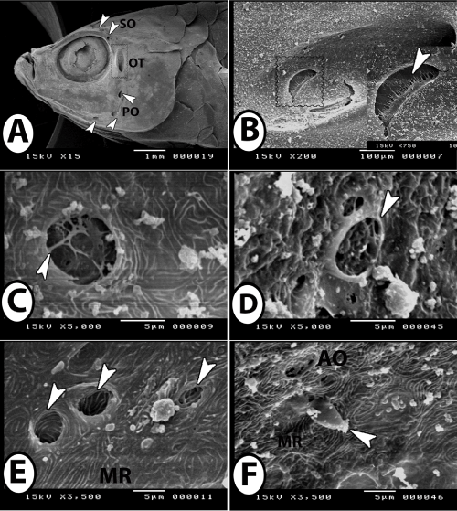
| Figure 9: SEM of lateral line system of guppy. A: The lateral line branch in the head into supraorbital (SO), otic (square) and preopercular (PO). B: Higher magnification of the square in Figure A showing ampullary organ (square, inserted figure) in a pit in the skin of the head that formed of receptor cells (arrowhead). C: Surface view of ampullary organ with attached cupula (arrowhead). D: Lateral view of ampullary organ (arrowhead) with alveolar-like appearance of the organ. E: Different size and shapes of ampullary pores (arrowheads). Note presence of fingerprint-like microridges (MR) of the surrounding epidermal cells. F: Superficial neuromast with attached cupula (arrowhead) that occur near ampullary organ (AO) among microridges (MR) of epidermal cells. |