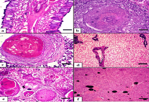
(b) Hepatopancreas of seabass showing large granuloma (H&E).
(c) Large granuloma in seabasshepatopancreatic tissue showing central
necrotic area encapsulated by fibrous connective tissue with some inflammatory cell aggregates(H&E)
(d) Hepatopancreas of seabass showing sever vacuolar degeneration of hepatocytes(H&E).
(e) kidney of seabass showing large records of granulomas replacing the renal tissue.(H&E).
(f) Spleen of seabass showing activation of melanomacrophage centers (H&E).