 |
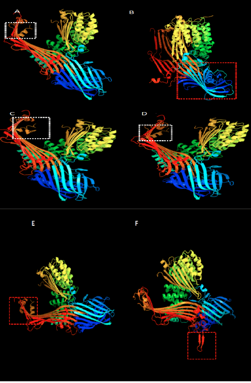
| Figure 5: The 3-D structure of Vtg in fishes from different species. Figure A, B, C, D, E, and F are the 3-D structure of Epinephelus lanceolatus, Ichthyomyzon unicuspis, Dicentrachus labrax, Clarias macrocephalus, Anguilla japonica and Danio rerio respectively. The location of helix; blue: helix1; green: helix 2; light green: helix 3; red: helix 4. |