|
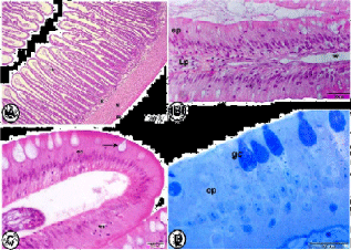
| Figure 2: Histological analysis and semithin sections of anterior intestine of grass carp. A: the wall of anterior intestine of grass carp was constituted of folded mucosa (m), submucosa (S), muscularis (M) and serosa (se). (Haematoxylin and Eosin). B: The mucosal fold of anterior intestine showing epithelium (ep) that consisted of simple columnar epithelium with goblet cells and lamina propria (Lp). Notice the presence of large lymphatic vessels (*). (Haematoxylin and Eosin). C: The simple epithelium (ep) of anterior intestine showing goblet cells (arrow) and wandering cells (wc). (Haematoxylin and Eosin). D: Semithin section of simple columnar epithelium (ep) of the anterior intestine of grass carp with goblet cells (gc). (Toluidine blue) |