 |
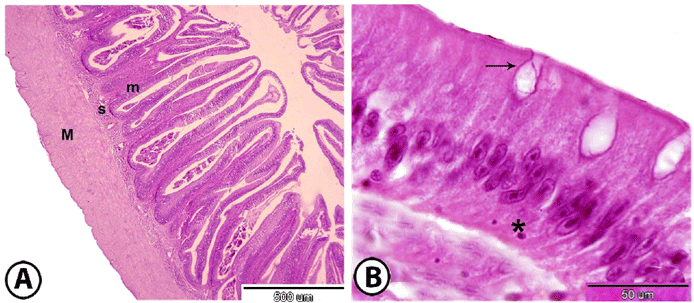
| Figure 1: Histological analysis of posterior intestine of grass carp stained with HE (A): the wall of posterior intestine of grass carp showing folded mucosa (m),submucosa (S) and muscularis (M). (B): Photomicrograph of the simple columnar epithelium of posterior intestineof grass carp with goblet cells (arrow). Note presence of lymphocytes in the basal part of the epithelium (asterisk). |