|
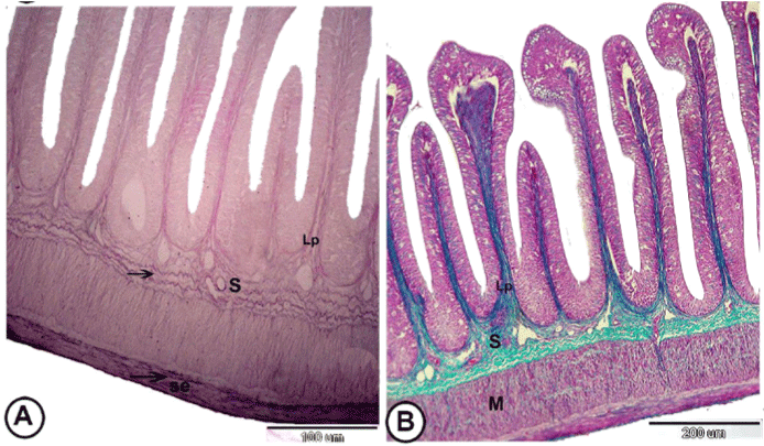
| Figure 3: The wall of the posterior intestine: (A) Photomicrograph of the posterior intestine of grass carp showing presenceof elastic fibers (arrows) in lamina propria (Lp), submucosa (S) and thick serosa (se). (Weigert’s Elastica, X 200). (B): Photomicrograph of the posterior intestine of grass carp showing laminapropria (Lp) and submucosa (S) stained green, while, tunica muscularis (M) stained red. (Crossmon’strichrome, X 100). |