|
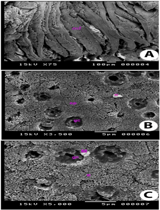
| Figure 5: Scanning Electron Microscopy of mucosa of posterior intestine: (A) Scanning electron micrograph of the posterior intestine of grass carpshowing mucosal folds (mf) separated by concavities. (B) Scanning electron micrograph showing the closely packed epithelial cells(ep) with goblet cells (gc). Notice the presence of mucous droplets (M) on the surface. (C) Scanning electron micrograph of the enterocytes (e) of the posteriorintestine of grass carp, covered by microvilli. Notice the presence of large goblet cells (gc) and mucous droplets (M). |