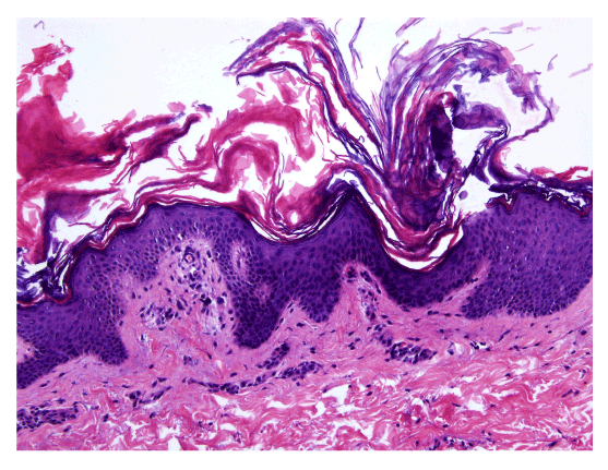
Figure 3:
Histological examination of the skin showed acanthosis with slight papillomatosis and ortokeratotic hyperkeratosis with a mild perivascular lymphocytic infiltrate on the upper dermis.