|
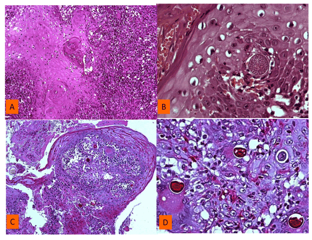
| Figure 2: A) Pseudoepitheliomatous hyperplasia of the epidermis and a dense infiltrate in papillary dermis (X10, haematoxylin-eosin stain); (B) Intraepidermal´s spherule with endospores (X40 haematoxylin and eosin stain); (C) granulomatous infiltrate in the dermis with spherules (X5 PAS stain) D) inflammatory granulation tissue with spherules inside of the multinucleate giant cells (X40 PAS stain). |