|
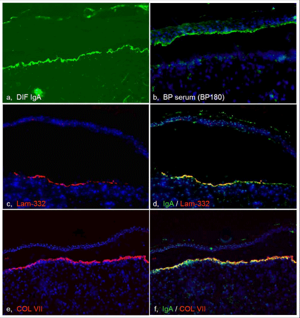
| Figure 2: Direct immunofluorescence showing linear IgA deposit at the dermal side of the separated BMZ (a). Immunostaining using the patients’ skin section revealed the epidermal BMZ labelling with a BP serum (targeting BP180 NC16a; b, green), but inversely showed the dermal BMZ labelling with laminin-332 (c, red) and type VII collagen antibodies (e, red). These two antigens overlay partially with the in vivo IgA deposit (yellow, d and f). DAPI was used for the nuclear staining (blue, b-f). |