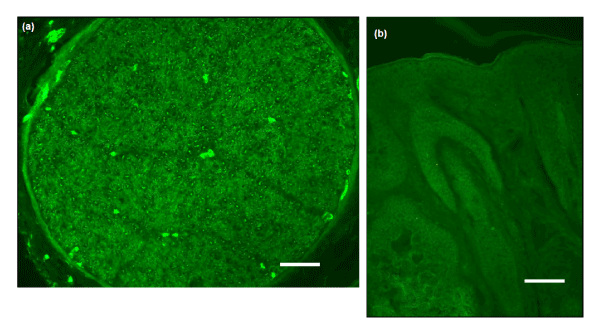
Figure 1:
(a) Positive control photomicrograph showing immunoreactivity in human spinal ganglion. (b) Negative control showing abolished staining after incubation with the primary antibody without IB4. Scale bars 40 μm.