|
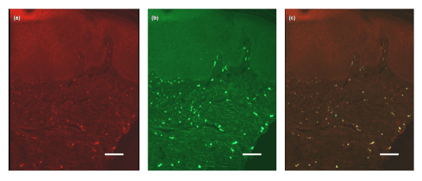
| Figure 3: (a) Double staining pictures showing IB4-labeled mononuclear cells (red colour) in the upper region of the dermis. (b) The same cells being tryptase-positive mast cells (green). (c) Colocalization seen with the double filter (orange-yellowish colour). Scale bars 40 µm. |