|
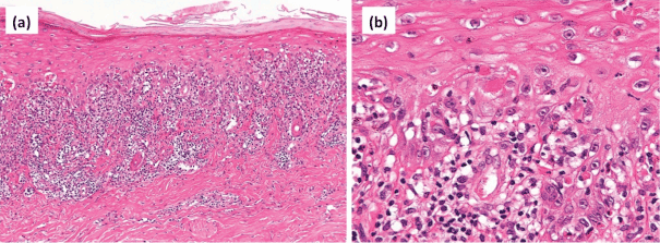
| Figure 2: Histopathological findings. (a) A lesion with lymphocytic infiltration extending into the papillae and into the epidermis. (b) Higher magnification showing satellite cell necrosis in the epidermis and lymphocytic infiltration. Hematoxylin and eosin, original magnifications (a) x100; (b) x400. |