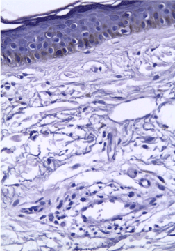
Figure 1c:
Mixed melasma; showing increased number of melanocytes and melanophages in the epidermal and dermal layers of the skin (H&E×400).