|
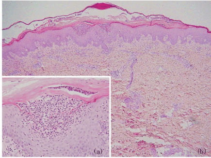
| Figure 2: Pathological findings of the skin lesions. (a) Biopsy of an abdominal pustule showed a spongiform pustule of Kogoj formed by neutrophils in the upper spinous and granular layers. (b) The epidermis showed parakeratosis and elongation of the rete ridges. The upper dermis contained an infiltrate of lymphocytes and neutrophils, migrating from the capillaries into the epidermis. |