|
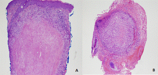
| Figure 1: Histologic spectrum of LMDF. A. Extensive caseous necrosis surrounded by a layer of histiocytes and multinucleated histiocytes with a peripheral rim of lymphocytes (40 × magnification). B. Predominance of cellular components with minimal central necrosis (40 × magnification). |