|
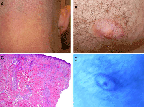
| Figure 4: A) Clinical exam notable for faint flesh colored papules on back of neck; B) Clinical exam notable for depigmentation of the nipples and areolae; C) Histopathologic exam of papules demonstrate prominent interfollicular granulomatous inflammation (Hematoxylin and eosin stain, 400x magnification); D) Wood’s light exam demonstrating enhancement of the nipples and areolae. |