 |
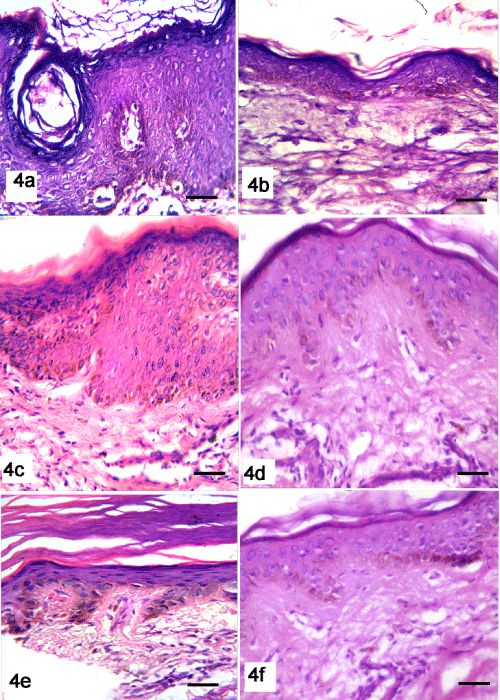
| Figure 4: Haematoxylin and eosin stained sections of group 2. seborrheic keratosis before peeling show hyperkeratosis, acanthosis and papillomatosis with a large horn cyst(4a), examination of skin after mix peeling reveal normal epidermis with increased epidermal thickness (4b). Actinic keratosis before peeling show hyperkeratosis, acanthosis and atypical epidermal cells (4c) while, after mix peeling skin show normal epidermal cells with increased epidermal thickness (4d). Solar lentigines before peeling show moderate elongation of the rete ridges with increased concentration of melanocytes and melanin pigments in the basal cell layer (4e), while skin after mix peeling show normal epidermis with decrease of melanin pigments in the basal cell layer (4f). (bar scale, 50 µm). |