|
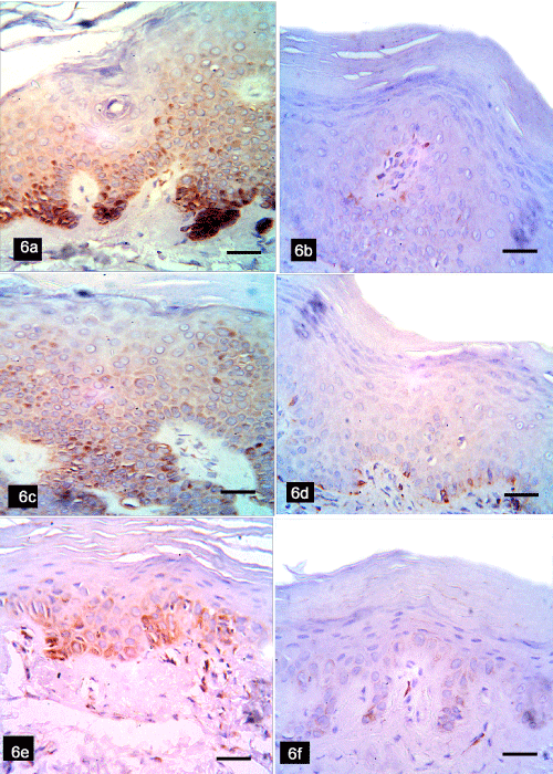
| Figure 6: Immunoperoxidase Bcl2 stained sections of group 2. Seborrheic keratosis before peeling reveal increased number of Bcl2 positive cells in basal and suprabasal layers of the lesion (6a), while after mix peeling show marked reduction in the number of positive cells limited to the basal cell layer (6b). Actinic keratosis before peeling show increased number of Bcl2 positive epidermal cells (6c), and after mix peeling show few positive cells limited to the basal cell layer (6d). Solar lentigines before peeling show increased number of Bcl2 positive cells in the basal and suprabasal cell (6e), while after mix peeling show moderate positive cells in the basal cell (6f) (bar scale, 50 µm). |