|
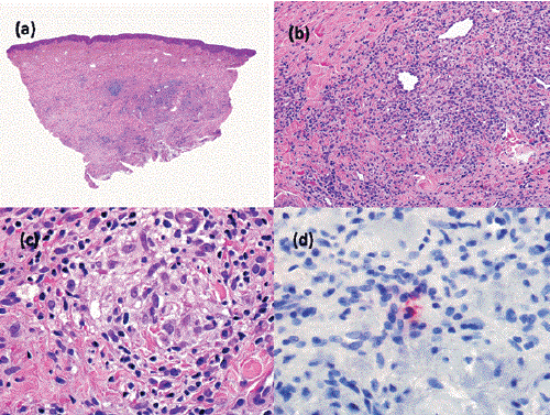
| Figure 4: Case 2. (A), (B) and (C), Hematoxylin & eosin stain. Like in Case 1, there was nodular dense inflammatory infiltrate composed of lymphocytes, plasma cells, histiocytes and neutrophils. (D), Immunohistochemistry showing Chlamydia trachomatis particles in lesional tissue. |