|
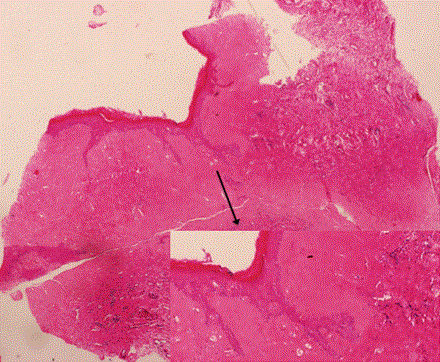
| Figure 1: Histopathology of a biopsy of affected vulval skin, stained with haematoxylin and eosin, showing hyperkeratosis, epidermal atrophy and over - hyalinised connective tissue in the underlying dermis. There is a mixed chronic inflammatory cell infiltrate below the hyalinised dermis and basal cell hydropic degeneration present at the basement membrane zone. |