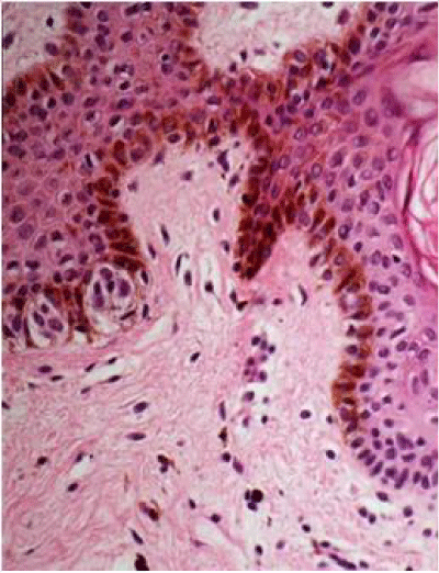
Figure 3:
Hematoxylin-eosin stain. Histopathological examination shows basal hypermelanosis and melanocyte nests in crests.