 |
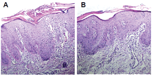
| Figure 3: Biopsy specimen from plaques of the purpose (case 1, A), and purpose’s father (case 2, B). Both Photomicrographs showing an epidermal hyperplasia with elongation of the rete ridges in a regular pattern, loss of granular cell layer, and overlying parakeratosis. Also present are blood vessels dilated and tourtuous, perivascular lymphocytic infiltrate, edema in the superficial dermis and presence of lymphocytes into the epidermis. Further, figure a presents intracorneal microabscesses and similar neutrophilic pustules in the spinosum stratum. In both cases findings are highly consistent with psoriasis. (Hematoxylin and eosin stain, original magnification x10). |