|
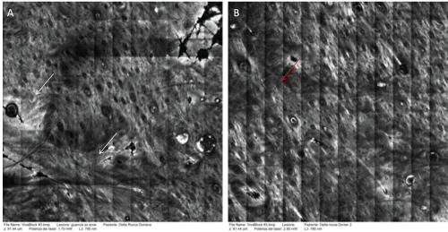
| Figure 4: a) Before the treatment, upper dermis of the scar was characterized by a network of fibers that appear thick and irregularly disposed (white arrows). b) After the treatment, we observe a more organized distribution of dermal fibers which were also seen as more fibrillar (red arrows). |