|
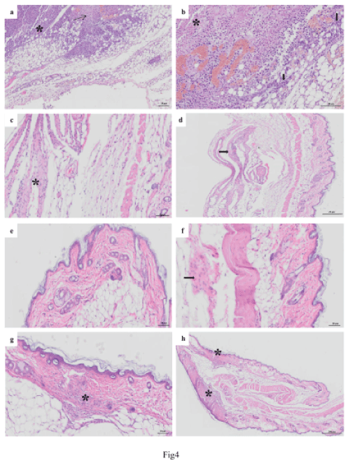
| Figure 4: Photomicroscopy of the subcutaneous tissue of C57BL/6 mice bearing B16/F10 melanoma, after 20 days of treatment. After the end of the treatment period, the animals were euthanized and the organs were collected for histological analysis. (a, b) Control tumor group; (a) Showing marked subcutaneous tissue by tumor infiltrating mononuclear cells (*). Note the presence of areas of necrosis (arrow); (b) Subcutaneous tissue showing invasion by tumor cells (melanocytes). These cells show on apoptosis and necrosis. With the presence of nuclear pyknosis. Presence of coagulation necrosis area (*). Note small area on the deepest portion showing preserved tumor cells (arrows). (c,d) Group treated with Etoposide; (c) Subcutaneous tissue showing healing process (increase of fibroblasts and collagen and presence of mononuclear cell infiltration (*); (d) Showing areas of skin healing process encompassing dermis and subcutaneous tissue (*). (e,f) RF followed by etoposide; (e) Skin showing the epidermis, dermis and skin appendages without histological changes; (f) Skin showing the epidermis, dermis, skin appendages without histological changes. Note area subcutaneous tissue showing healing process (increase of fibroblasts and collagen - arrow). (g,h) Etoposide followed by RF; (g) Showing skin healing process area with increasing fibroblast, collagen and mononuclear inflammatory cells (*); (h) Skin showing the epidermis, dermis and subcutaneous tissue attachments without histological changes. |