|
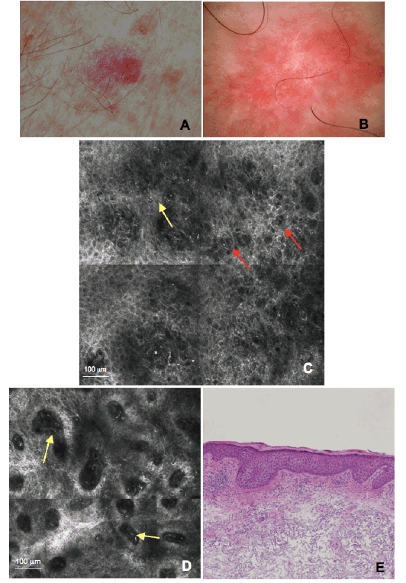
| Figure 2: Clinical photograph of a red scaly plaque of the arm (A). This lesion shows on dermoscopy (B) features suggestive of SCC such as vascular pattern characterized by vessels appearing as red dots. CM mosaic image (C, 1 x 1 mm) at the level of the spinous granular layer shows atypical honeycomb pattern with pleomorphic cells and broadening of the intercellular spaces (red arrows), and small bright cells suggestive of inflammatory cells (yellow arrow). CM image (D, 0,75 x 0,75 mm) at the level of the DEJ shows dilated, round blood vessels within dermal papillae (yellow arrows). On histopathology (E), the lesion proved to be a SCC in situ. |