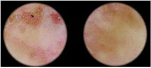
Figure 2:
Dermoscopic examination shows an atypical vascular pattern with residual pigmentation and milky red areas.