|
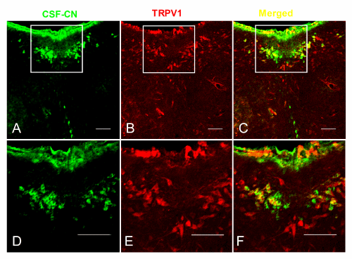
| Figure 3: TRPV1-expression in the normal SD rats. A: CB-HRP positive neurons (green). The cell bodies of dCSF-CNs were in the brain parenchyma and the processes were extended into CSF. B: the same section showing TRPV1 positive neurons (red). C: same section showing CB-HRP/TRPV1 double-labeled neurons (arrow, yellow). D-F are showed the enlargement of the rectangle in A-C. Scale bar: 100nm. |