B. Case 1. Postoperative plain CT demonstrating a small hemorrhage in the left thalamus after stereotactic biopsy (arrow).
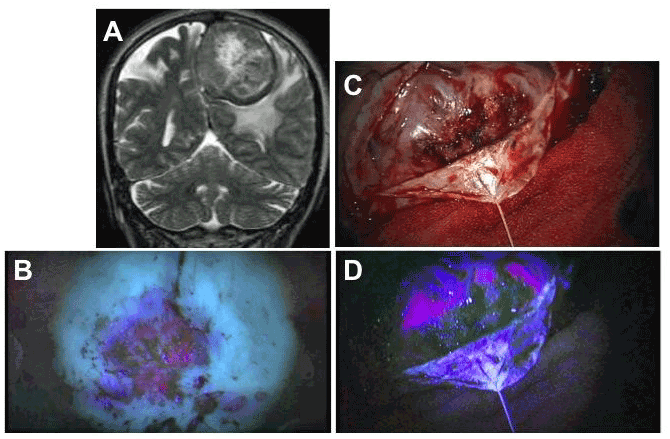
C. Case 2. Preoperative, axial, T2-weighted, MR image demonstrating a massive, diffuse, pontine glioma.
D. Postoperative, axial, T2-weighted, MR image demonstrating a high-intensity area after stereotactic biopsy (arrow).