|
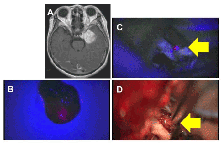
| Figure 2: A,B. Case 1. Tumor specimen of malignant lymphoma under white light illumination (A) and under a violet-blue light (B). One pair of tumor specimens at the left shows charcoal-red fluorescence; in contrast, the negative control of subcutaneous tissue at right shows no fluorescence (B). C,D. Case 2. Tumor specimen of pontine glioma under white illumination (C) and under a violet-blue light (D). One pair of tumor specimens at left shows slight charcoal-red fluorescence; in contrast, the negative control of subcutaneous tissue at right shows no fluorescence (D). |