|
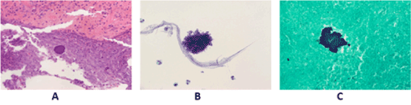
| Figure 2: (A) Histological Examination of brain abscess revealing actinomycotic colony surrounded by necrosis and inflammatory infiltrate (hematoxylin and eosin stain, ×20). (B) Photomicrograph of aspirate showing mixed inflammatory cell exudates and fluffy colonies. The actinomyces organisms radially arranged filamentous bacilli (hematoxylin and eosin stain, ×40). (C) Cell block section showing positive silver impregnation of actinomycotic colonies(Grocott-Gomori methenamine-silver nitrate, ×40). |