 |
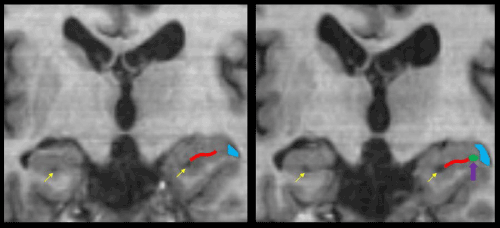
| Figure 1: Hippocampal sulcal cavity (purple arrow) and vestigial hippocampal sulcus (yellow arrows and red lines) enlargement in a patient with moderate TBI at Time 1 (left) and Time 2 (right). Note that the lateral ventricles and inferior horn of the lateral ventricles (IHLVs, colored in blue) also become larger. The MRI scanner used was a General Electric Signa-Echospeed 1.5 Tesla (GE Healthcare, Milwaukee WI), using an eight channel head coil. The image presented was from the high-resolution 1 mm isotropic T1 weighted, three-dimensional IR prepped radio-frequency spoiled-gradient recalled-echo (3D IRSPGR) sequence (TI/TR/TE=12/300/5,TI, FA=20, slice thickness=1mm no gap, matrix=256 × 256) with images acquired in the axial plane utilizing a 25cm field of view. This pulse sequence acquires high-resolution structural data, which is the case of T1-weighted, implies a slice thickness and in-plane dimensions of no more than 1mm, which we have found necessary to view HSCs. Although acquired in the axial plane, the isotropic acquisition allows the data to also be viewed from other planes, such as sagittal and coronal. In our experience, the optimal plane for viewing HSCs is coronal. We have also found that HSCs are best viewed in native (raw) space as normalization may squash HSCs rendering them to no longer be visible. We urge caution when filtering as it usually blurs HSC boundaries. |